


 Share
Share


Brand: |
none |
Address: |
China |
Min.Order: |
none |
Supply: |
none |
Valid until: |
Long Term |
Type: |
Array |
Product Details
1.15-inch high resolution LED monitor 2.standard PC LED backlit silicon gel keyboard
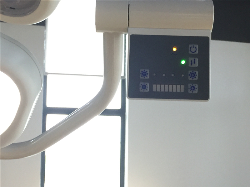
3.one key regulation, easy operation
4.rechargeable built-in battery
5.abundant peripherals: DICOM3.0, VGA, video out, USB etc
1. Detailed Product Description
A.Imaging optimization technology;
B.Compound enhance technology
C.Imaging processing technology
D.Imaging optimization technology
E.Compound enhance technology
F.Multi-beam parallel processing technology
G.Tissue harmonic imaging

2. Displaying mode
B, 2B, 4B, B/M, M
B/C, B/C/D, B/D CFM, PRF
PW, velocity, power (direction), histogram
Triplex / Duplex
3. Optional Probe
Wide band multi-frequency
Electronic convex probe
Trans-vaginal probe
Linear probe
Rectal probe
Micro-convex probe
4. Clear ultrasound images
Probe information , display mode, the depth of focus , the dynamic range of standard body , the probe position mark , sound power ,
patient information, medical institution name, the measurement value, time and date , the scale , the scanning direction ,gamma curve ,
the probe current work Frequency, frame rate , B mode overall gain , C mode overall gain , D model of the total gain , menus, comments,
gray belt, puncture guide line , PRF, wall filter , blood-related , cumulative frequency , TI heat index , MI machinery index
Storage function:
Probe parameter storage
Image Storage
Cine storage
Measurement results are stored
Report storage
Grayscale :256 gray
Interface Language:Optional English
Trackball function:
According to the working status and menu execution , completion of various operating functions
Control multiple electronic device, position the cursor , complete the measurement Function
5. Technical Parameters
Displaying mode: | BB,4B,BM,M,PW,B/C,B/C/D,B/D,duplex, triplex, part zoom, CFM, CPA |
Signal processing: | Full-digital beam forming, dynamic filter, space-time filter, dynamic real time receiving focusing, RDA, DRA, spectral processing, CFM processing, gray scale 256, receiving signals dynamic range≥150, All digital, real-time dynamic focusing, dynamic aperture in all fields |
Image processing: | Image persist THI Speckle-reduction Color coder, Power adjustable Multi-frequency Smoothing function Edge enhancement Image optimizing disposal One-key optimization Probe mark Image conversion Up/down conversion Left/right conversion Doppler Sound output volume adjustable Wall filter adjustable Base line adjustable Sampling frame adjustable Area changeable(angle changeable) Spectrum sampling volume adjustable Spectrum sampling volume angle adjustable PRF adjustable |
Gain control: | TGC, PW Gain, CFM gain |
General measurement: | B mode-distance, circumference, area, volume, ration, angle, % stenosis, histogram M mode- distance, time, velocity, heart rate |
OB packages: | EDD table :GS ,BPD, CRL,FL,HL,TAD,LV,OFD,NT,AC,HC,AFI GA and EDD calculated by LMP, BBT calculate GA and EDC, Fetal physiological score, Fetal growth curve OB measuring and calculation report |
Gynecological packages: | Uterine measurement (Cervical length, endometrial thickness, uterine volume) Gynecological measuring and calculation report |
Urology packages: | left/right kidney measurement vesicant urinary /After voiding bladder measuring Urology measuring and calculation report residual urine’s measurement |
Anthology packages: | Prostate Testis Anthology measuring and calculating report PPSA,PSAD calculation |
Peripheral vascular measurement | stenosis, Diameter stenosis rate, Peripheral vascular measuring and calculating report |
Small parts Measurement | Thyroid,Mammary gland,Mass,Small parts measuring and calculating report |
| Multiple Births measurement | |
| Orthopedic surgery measurement | Left/right hip |
Cardiac Measurement Package | Heart rate, Valve speed, LV, aortic, mitral, ventricular |
Doppler Measurement | Time Heart Rate Velocity General Measurement Doppler automatic measurement Doppler tracing (auto/manual) |
Body mark | Abdominal,Anthology,Cardiology,Gynecology,Musculoskeletal,Obstetrics,Pediatrics Small parts,Urology |
| Comment and display | Real time clock,Comment in image area,Scanning density,TI value,MI value |
Cine loop | Automatic cine loop frames, Replay of stored screen frames, Selection of replay speed, Frame replay, Replay stored files, Cancel, replay, pause control |
Image storage format | BMP,JPEG,PNG,DICOM, |
Image storage size | ≥320G |
Installed work station | OB report, GYN report Urology report Cardiac measurement - LV report Cardiac measurement - mitral report Cardiac measurement - Ventricular report Small parts measuring report Peripheral vascular measuring report Diagnostic template Printing report |
Input/output ports: | VGA USB port DICOM port(network port) RS232 Laser printer (option) Video printer (option) |
Standard Configuration | Main Unit, 15 inch high definition color LCD, 3.5MHz multi-frequency, User’s Manual, 60G(increasable) Image storage size |
Option | Probes, trolley, video printer, laser printer, puncture guide standard |
Customer Reviews total 0 items